

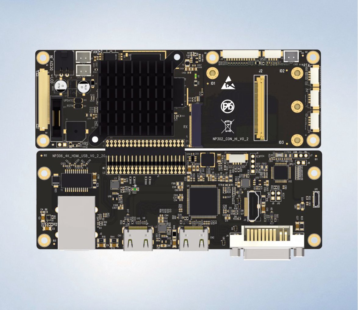
The sensor: where contrast is decided
A 4K endoscopic camera module starts with a large-format CMOS sensor. In the dark, humid, reflecting world of the surgical cavity, sensor size and pixel architecture matter more than headline resolution. Larger pixels gather more photons per frame, which translates directly into lower noise and cleaner shadow detail when the scope is pulled back for orientation. Dual-gain architectures add a second lever: they read the same scene at two conversion gains and merge the results, keeping highlights intact while lifting the darkest tissue planes out of the noise floor.
It is worth being precise about what "large" buys you. Photon count scales with pixel area, and shot noise scales with the square root of that count — so a pixel with four times the area carries roughly twice the signal-to-noise ratio, not four times. That is why two sensors with identical resolution but different optical formats produce visibly different images in the same scope, under the same light, at the same gain. The datasheet number that predicts this is not the megapixel count; it is the pixel size and the sensor's quantum efficiency in the wavelengths your light source actually emits.
Why "4K" is the least informative number on the datasheet
4K describes the delivery format, not the imaging quality. A 3840 × 2160 pipeline fed by an under-sized sensor, a narrow-bandwidth link and an uncalibrated monitor produces 8.3 million soft, noisy pixels — technically 4K, clinically no better than a good 1080p chain. Resolution is only real when every stage in front of it can resolve: the optics must deliver modulation at those spatial frequencies at your working distance, the sensor must sample them without aliasing, the link must carry them without compression artefacts, and the display must show them without scaling.
The practical consequence: when a vendor says "4K", the next question is 4K of what. Ask what the limiting resolution of the complete chain is at 20 mm working distance, in line pairs per millimetre, through the actual scope that ships with it. A vendor who can answer that has measured the chain. One who quotes only the panel format is selling you the easy number.
The ISP: where colour is decided
Raw sensor data is not an image a surgeon can act on. The image signal processor performs demosaic, white balance, flare compensation and detail enhancement — all under illumination that shifts between LED white light and laser-based fluorescence. The hardest engineering problem is channel alignment: when a module drives both a white-light stream and an ICG fluorescence stream, the two must be fused at pixel level. A misaligned overlay of half a millimetre is the difference between identifying a bile duct and cutting it.
Alignment failures rarely come from the sensor. They come from everything around it: a filter placed in front of the sensor shifts the optical path slightly; the two illumination wavelengths focus at marginally different planes; a mechanical tolerance stack in the coupler moves the field between modes. The fix is calibration as a system, not as parts — aligning the fused overlay through the assembled scope, across the full working-distance range, and verifying it after every reprocessing-relevant service event. It is unglamorous work, and it is exactly the work that separates a fused image you can operate on from a picture-in-picture toy.
The bandwidth nobody budgets for
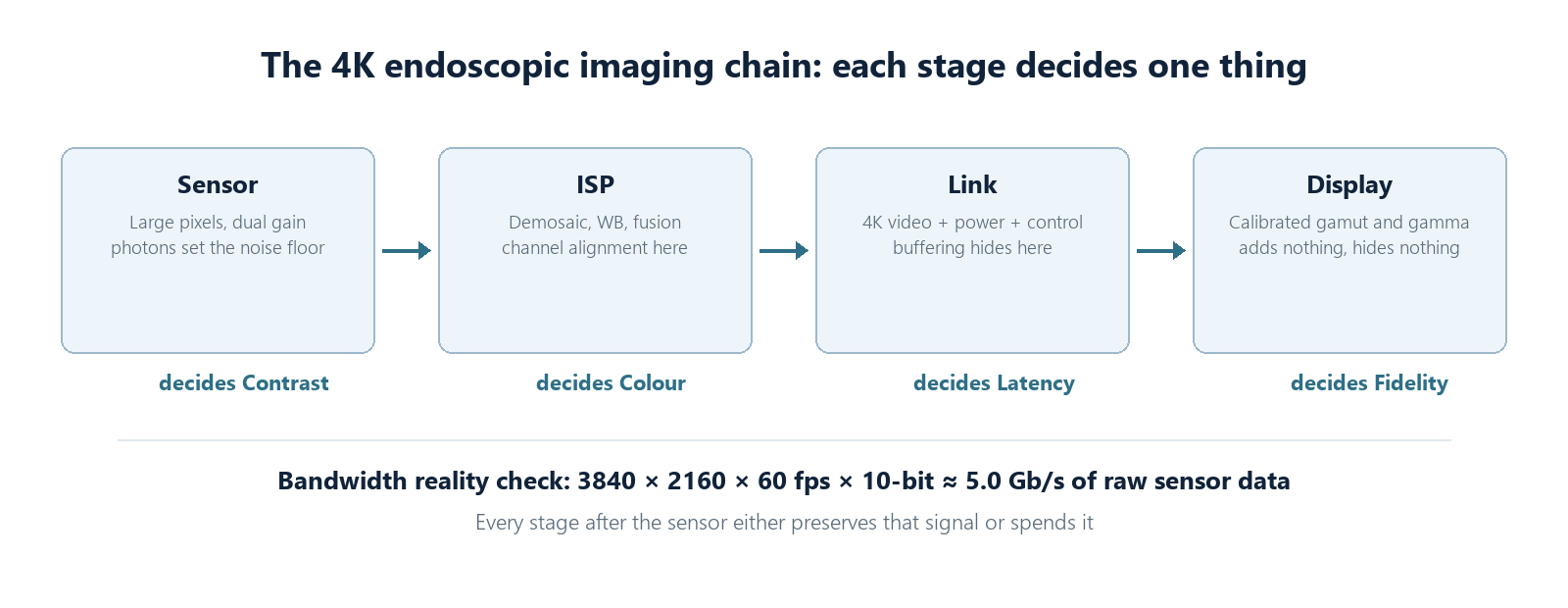
Run the arithmetic once and it changes how you read datasheets. 3840 pixels by 2160 lines at 60 frames per second is a quarter of a billion pixels every second; at 10 bits per channel of raw Bayer data that is roughly 5 gigabits per second before any overhead. Something has to happen to that stream: either the link carries it compressed, or it carries it uncompressed and pays for the copper, or the pipeline processes it locally and sends the result. Every choice has a failure mode — compression latency and blocking, copper cost and stiffness, processing heat in a camera head small enough to hold.
This is why two "4K60" systems can behave nothing alike. One ships 5 Gb/s of honest pixel data down its cable; another ships a compressed approximation of it. The compression is not visible on a static test chart. It becomes visible the moment tissue moves, when every block-boundary and every dropped bit lands on the anatomy you are dissecting along.
The link: where latency is decided
Once the image exists, it has to travel — through a cable rated for repeated sterilization and mating, to a monitor the surgeon actually watches. Every frame the pipeline buffers adds delay between hand movement and on-screen result. A well-designed single-coax link carries 4K video, power and control on one cable with zero perceptible latency, which is why the link stage, not the sensor, is where most "fast" systems quietly become slow.
Latency: a budget, not a vibe
End-to-end latency is the sum of small leaks: sensor readout, ISP processing depth, codec buffering, link serialization, display pipeline. Individually each sounds acceptable — a frame here, half a frame there. Summed, they decide whether the image feels welded to the hand or dragging behind it. At 60 fps a frame is 16.7 ms; four frames of accumulated buffering is 67 ms, which is around the threshold where precise manipulation starts to feel wrong to a trained hand.
The reason this keeps happening is that latency is nobody's headline spec. Sensor vendors quote sensor latency, processor vendors quote processing latency, and the sum appears on no page of any datasheet. The only defence is measurement: a metronome or a flashing LED filmed together with its monitor image, one frame divided by the other. If a vendor will not do that test in front of you, assume the number is worse than theirs.
The display: where nothing should be added
The final stage should be honest. A surgical monitor with the right colour gamut and calibration adds nothing and removes nothing. Systems that look impressive in a demo room but fall apart in the OR usually failed here — or earlier — in places a datasheet never mentions.
The display stage has its own quiet failure modes. A consumer panel rescales every input to its native grid, softening the very detail the chain worked to preserve; its factory gamma targets a living room, not a shadow-rich cavity; and its default sharpness processing manufactures halos around vessels. A surgical display keeps the chain's resolution end to end, holds a stable gamma suited to endoscopic viewing, and leaves the image alone. The best compliment a display can earn in an OR is that nobody comments on it.
What to compare, in what order
When evaluating complete chains, the productive order is: contrast at working distance first, then colour fidelity against a reference target under the actual light source, then white-light/fluorescence overlay accuracy, then end-to-end latency, and only then resolution charts and panel format. The first four are where systems differentiate; the last two are where brochures live. A chain that wins on the first four will look better in the OR regardless of what its spec sheet says about the fifth.
Related reading: the camera control units and imaging modules this chain is built from, and our notes on FPGA versus SoC processing backends.