

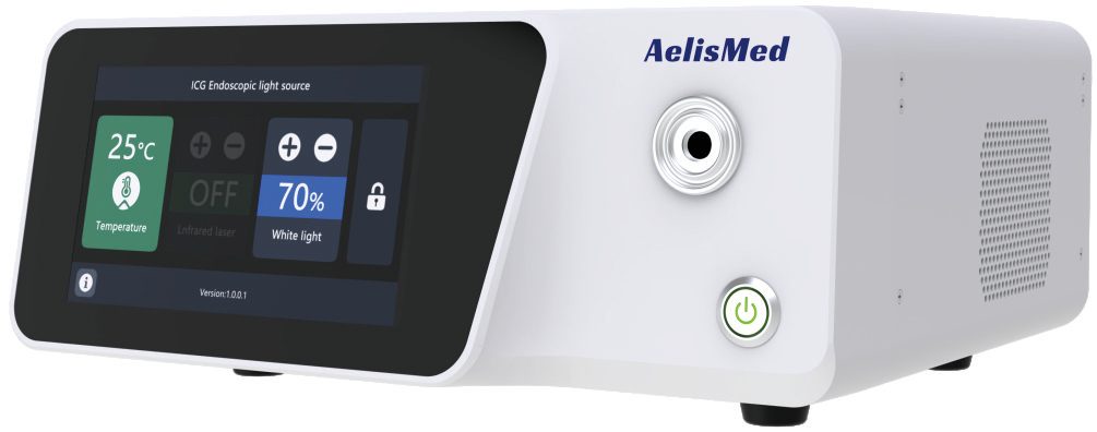
How ICG imaging works
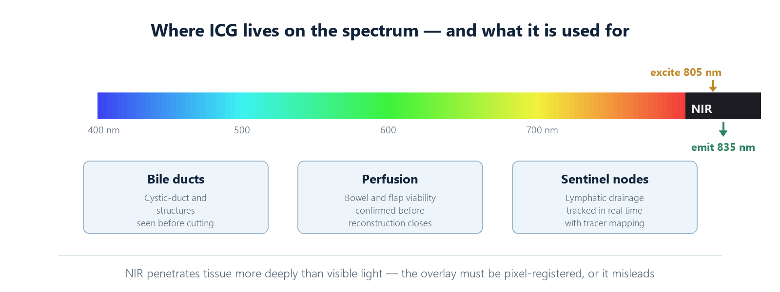
Indocyanine green binds to plasma proteins and fluoresces in the near-infrared: excitation around 805 nm, emission around 835 nm. Because NIR penetrates tissue more deeply than visible light and is invisible to the naked eye, the surgeon sees a real-time perfusion map overlaid on the standard white-light view — vessels, bile ducts, ureters and perfusion boundaries made visible.
The physics that makes this useful also makes it unforgiving. The fluorescence signal is faint — a small fraction of the excitation light returned at a shifted wavelength — and everything upstream of the surgeon's eye must be built to preserve it: an excitation source stable enough to produce repeatable signal, optics that block the excitation wavelength from leaking into the detection path, a sensor genuinely sensitive at 835 nm, and a fusion engine that places the fluorescence exactly where the corresponding white-light anatomy sits.
Where it earns its place
The established workloads explain why ICG has spread so quickly. In cholecystectomy, a green-tinged biliary tree turns the critical view of safety from an inference into an observation. In colorectal surgery, anastomotic perfusion can be assessed before the anastomosis is created, putting perfusion data where the leak risk actually lives. In reconstructive work, perforator mapping and flap-early-perfusion checks happen in the same session as the raise. And in lymphatics, tracer mapping makes drainage visible without ionising radiation.
What these share is a decision that is currently made on indirect evidence. Fluorescence does not make the decision — it upgrades the information the decision is made from. That framing matters when evaluating systems: the question is never "does it glow" but "is the glow where the anatomy is, at an intensity that means something".
Laser or LED: the excitation question
Excitation sources divide into laser diodes and LEDs. Lasers deliver a narrow, stable, intense 805 nm line with predictable output across the rated lifetime, which keeps fluorescence signal strength repeatable between cases. LEDs are cheaper and simpler but drift with temperature and age, which shows up as inconsistent signal between the first case of the day and the fifth. For perfusion-critical decisions, stability wins.
Drift is sneaky because the eye compensates. A slow output decay reads as "the patient's perfusion looks a bit weaker today", not as "the light source has aged". The systems that avoid this trap either stabilise the source actively or expose its output on screen so the operator can tell signal change from illumination change. Ask which yours does; if the answer is neither, every quantitative claim the system makes is on sand.
Dose and timing belong to the surgeon
The imaging chain's job ends where the pharmacology begins. Dosing regimens, injection timing and the interval between bolus and observation differ across procedures and institutions, and they change what the image shows: visualise too early and the signal is background, too late and the interesting phase has passed. A well-designed system supports this rather than fights it — with exposure and gain controls that respond predictably, a timeline that lets the team replay the arrival phase, and recording that captures the dynamic study rather than only its endpoint. What it should never do is hide the dose-and-timing dependency behind an automatic mode that silently renormalises every frame.
What the sensor side must do
Capturing faint NIR emission while simultaneously delivering a full-quality white-light image is a balancing act. The sensor needs genuine near-infrared sensitivity — not a heavily filtered consumer part — and the two image streams must be aligned at pixel level before fusion. An overlay that is "roughly" registered will still impress in a demo and mislead in the OR.
Quantitative, or just a glow effect
The difference between a measurement and a mood is repeatability. A fluorescence image becomes clinically useful when intensity maps to something stable: the same perfusion state produces the same brightness, on the same system, between cases. That requires a calibrated excitation source, a defined gain structure, and a display convention that does not renormalise away the very differences being judged. Systems that auto-expose in fluorescence mode are actively destroying the information the surgeon is trying to read — every frame is re-scaled against itself, and a improving or worsening perfusion gradient disappears into the gain loop.
The practical standard to demand: absolute or at least consistent presentation of signal, side-by-side white-light and fluorescence views available alongside any fusion, and documentation of the gain state in the recording. Anything less is a night-light with a brand.
Questions worth asking a vendor
- What is the overlay accuracy between white-light and fluorescence channels, in pixels, across the working-distance range?
- Is the excitation source output stabilized, and over what lifetime?
- How is fluorescence gain presented — normalized, or absolute?
- Can the system display both channels side-by-side, not only fused?
- Does the recording preserve the dynamic study with gain state, or only the fused image?
Fluorescence imaging earns its place in the OR when it is quantitative, stable and precisely registered. Everything else is a glow effect.
Related reading: our ICG fluorescence imaging modules, the light source modules behind the excitation side, and the anatomy of the full 4K imaging chain these modules plug into.