

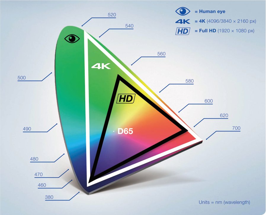
Most endoscope image complaints trace back to five artifacts: moiré, washout, lag, colour cast and low-light noise. Each has a distinct cause — and a fix that is either optical, electronic or free. A field guide to identifying which one you are looking at.
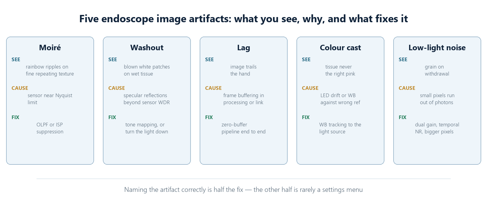
The value of naming them precisely is that the five fail differently. Moiré is geometry. Washout is dynamic range. Lag is buffering. Colour cast is calibration. Low-light noise is physics. Each points at a different stage of the chain, and each has a different owner: the optical designer, the ISP engineer, the link architect, the clinical engineer with a reflectance standard, or the sensor itself. "The image looks bad" sends everyone in the wrong direction at once; "the image shows moiré on suture material at close working distance" usually sends exactly one person.
1. Moiré — the wavy interference pattern
Fine repeating structures in tissue or sutures beat against the sensor's pixel grid, producing rainbow ripples that are not in the patient. Cause: the sensor is resolving detail close to its Nyquist limit without an optical low-pass filter. Fix: optical or ISP-level suppression; sometimes simply changing working distance removes the beat frequency.
Moiré deserves respect rather than a blanket fix, because the cure can cost more than the disease. An optical low-pass filter blurs everything by design — it trades a little genuine resolution everywhere to remove the interference in the rare frame that needs it. ISP suppression is more surgical but has to be conservative: aggressive moiré filtering and fine vessel texture live in the same spatial-frequency neighbourhood, and a heavy hand deletes the diagnostic detail along with the ripple. The honest approach is mild suppression plus awareness — the artifact is information about where the chain's limits are.
2. Washout — blown highlights on wet tissue
Specular reflections from wet tissue and instruments saturate entire regions, hiding the anatomy underneath. Cause: insufficient highlight handling in the sensor readout or ISP tone mapping. Fix: highlight-recovery tone mapping, wider dynamic range readout, or — cheapest of all — adjusting the light source output before reaching for the settings menu.
The hierarchy of fixes matters because each level costs more than the last. Light-source discipline is free and solves the majority of cases: the specular highlight that swamps a region is usually an illumination choice, not a sensor limit. Tone mapping recovers what illumination discipline cannot, and modern dual-gain readouts widen the window at the source. What does not work is cranking local contrast or sharpening to "bring back" saturated regions — the information is not there to sharpen. A saturated pixel is a statement that the scene exceeded the capture window, and no menu setting travels back in time.
3. Lag — the image that follows the hand
Move the scope; the image catches up a beat later. Cause: frame buffering somewhere in the chain — usually in processing or transmission, not the sensor itself. On a 60 fps system, four buffered frames add 67 ms: enough to make precise manipulation feel like driving through syrup. Fix: a zero-buffer pipeline; measure end-to-end latency, not sensor latency.
Lag is the artifact most often misattributed. Because the sensor is the component people think of first, and because sensor readout is rarely the bottleneck, teams spend weeks tuning acquisition while the delay hides in a processing buffer two stages later. The measurement discipline that settles it is simple and should be standard acceptance procedure: film a millisecond clock on the monitor next to the real object, and divide the difference by the frame time. Every component vendor's latency claim becomes irrelevant the moment that one number exists.
4. Colour cast — the tissue that is never the right pink
A global green, magenta or blue shift that survives every white-balance reset. Cause: illumination drift with LED age or temperature, or a white balance calibrated against the wrong reference. Fix: continuous white-balance tracking referenced to the actual light source, and recalibration against a reflectance standard rather than a white gauze.
The reference standard point deserves its own sentence, because it is the most common process failure in the field. White gauze is not white in any calibrated sense — it fluoresces slightly, its weave shades under directional light, and its age changes its reflectance. A chain white-balanced against gauze inherits every error the gauze carries. A reflectance target costs less than the time wasted chasing phantom colour shifts, and it converts colour disputes from opinion into measurement.
5. Low-light noise — the grain that appears on withdrawal
Pulling back for orientation drops light levels and the image dissolves into grain. Cause: small pixels running out of photons. Fix: larger pixels, dual-gain readout, or temporal noise reduction that respects tissue motion. Digital brightness boost alone amplifies the noise with the signal.
The withdrawal case is worth understanding because it is the noise condition surgeons actually complain about, not the close-work condition where light is concentrated. As the scope retreats, the same LED output spreads over a wider field, per-pixel illumination falls, and the shot-noise floor rises relative to signal. Systems handle this at the source — bigger pixels, dual conversion gain, an aperture strategy in the optics — or at the ISP, where temporal noise reduction that tracks tissue motion can buy real visibility without the smearing that makes vessels vanish. What to distrust is the digital gain slider: it brightens the noise exactly as much as the anatomy.
A five-minute triage routine
When the image looks wrong, the fastest path is sequence, not intuition. Set the working distance and illumination to the standard condition and ask which artifact is present by name. Change one variable at a time — light output first, working distance second, gain third — and note at which step the artifact moves. If nothing changes it, the cause is upstream of optics and the calibration or pipeline team should look; if a free variable fixes it, log the condition, because the fix that "works" without being understood becomes tomorrow's mystery. Five disciplined minutes routinely saves an afternoon of finger-pointing.
None of these artifacts require replacing a system. All of them require knowing which one you are looking at — which is the point of writing their names down.
Related reading: the imaging modules these pipelines run in, how a thousand reprocessing cycles gradually produce some of these artifacts, and the anatomy of the full 4K imaging chain.